|
Radiation Dosimetry
Aim Development of a comprehensive medical radiation dosimetry system
Background Advanced radiotherapy techniques produce radiation dose maps with high dose modulation and tight gradients. Difficulties in the dosimetric verification of these new complex treatment methods using existing dosimeters has led to the need for a new generation of fast responding real time dosimeters with submillimetre precision. In most clinics, routine quality assurances for machine output are generally in place to ensure the constancy of day to day radiation treatment delivery. However, as radiation therapy treatments becomes more and more complex, the role of supplementary dosimetry such as patient specific and in-vivo dosimetry are gradually gaining importance. AAPM TG 40 recommended that clinics should have access to TLD or other in-vivo system in order to prevent major treatment errors. To that end, thermoluminescence dosimeters (TLDs) have been widely used for in-vivo dosimetry. In diagnostic radiation imaging, radiation dose measurement has never been more important. This is brought on by the advancement of medical imaging technologies such as the multislice Computed Tomography (CT) and biplanar angiography. The inadvertent increase of patient dose with these imaging modalities needs to be reassessed. Over the past decade, researchers at the University of Malaya have been monitoring the radiation dose to patients receiving various diagnostic imaging procedures in Malaysia. These works were primarily based on TLD, compliment by other dosimeters such as film dosimetry. Recently, a new type of TLD was developed using aluminium oxide as the substrate. This material has shown to response to the stimuli of visible light, leading to the development of optically stimulated luminescence dosimeters (OSLD). This new technique is highly sensitive to low dose and allows multiple readouts. The ease of use of these detectors has seen the adoption of this technology by the Radiological Physics Centre (RPC), United States.
Optically Stimulated Luminescence (OSL) Dosimetry Optically stimulated luminescence dosimeters (OSLDs) are plastic disks infused with aluminium oxide doped with carbon (Al2O3:C) crystals. These disks are encased in light tight plastic holder. This type of crystals when exposed to ionising radiation are able to store radiation energies through excited electrons that fall into deep traps, whereas the readout mechanism utilises light exposure. The OSLD has been characterised and used in the quality assurance of radiation therapy Compared to. TLDs, the OSLDs are a relatively new practical dosimeter which allows multiple readouts without deterioration of dose information that is major advantage in comparison with TLD. However OSL has stronger energy dependence in comparison with LiF . More basic work is required to understand the full capacity and the use of this dosimeter. Clinical applications and implementation of this dosimetry for in-vivo dosimetry is still not widely done. Hence, there are opportunities to develop and promote of the use of this detector in this area. The use of OSLD in the diagnostic imaging dose measurement is still at its infant stage. To the best of our knowledge, early characterisation of the energy and angular dependence of the OSLD in the diagnostic energy range (20 kV - 200 kV) has only been reported by two groups. We proposed to study of the OSLD in an anthropomorphic phantom for accurate dose measurements. OSLD has been primarily used as a passive detector which allows detector readout at a later time after irradiation. However, as time is a crucial factor in many clinics, a detector having real time readout ability will be very attractive and useful in the quality assurance and patient dose verification. We proposed to develop such a real time readout system based on fibre optic technology.
Semiconductor Dosimetry Semiconductor radiation detectors play an important role in radiation dosimetry. This is due to their many advantages such as small size, excellent energy resolution, and ability to be integrated with fast electronics readout allowing real time dose measurements. Semiconductor radiation detectors have been used side by side with the ionisation chamber in the quality assurance of radiation therapy beam. Semiconductor diodes and TLDs have been widely used for in-vivo dose measurement such as in the treatment verification of total body irradiation (TBI) and total skin irradiation (TSI). However, with proper calibration, semiconductor detectors can have better spatial and dose accuracy than TLDs. Conventional diodes are packaged with an epoxy bubble-encapsulated build up material. This precludes the use of these detectors for skin dosimetry since skin depth is defined at the depth of 0.07 mm. MOSkin dosimeter developed by the Centre for Medical Radiation Physics (CMRP), University of Wollongong is based on MOSFET technology. The design of the detector is particularly suitable for skin dosimetry since it has a water equivalent depth (WED) of 0.07 mm. |
|
|
Areas of research · Radiochromic film dosimetry · Optically stimulated luminescence (OSL) dosimetry · Semiconductor dosimetry o In-vivo dose measurements: Breast skin dose during radiotherapy treatment o Eye lens dose measurement o Peadiatric CT dose measurements |
|
|
|
|
|
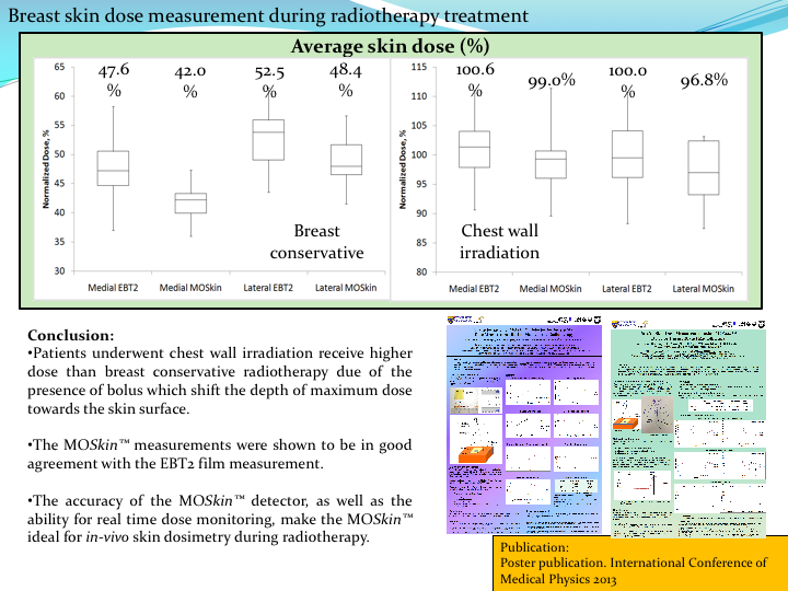
Breast skin dose during radiotherapy treatment
Determine skin dose for breast radiotherapy - to ensure sufficient dose to the treatment volume without excessive skin reaction. Current treatment planning systems are unable to accurately calculate the dose in the build-up region. Objective: To measure actual skin dose through in-vivo skin dosimetry.
|
|
|
Eye lens dose measurement
Threshold dose to induce non-cancer tissue reaction in eye lens - 0.5 Gy. Long beam-on time during neuro-interventional procedures may deliver doses in this magnitude to the patient’s eye lens. Objective : To determine eye lens dose during neuro-interventional procedures
|
|
|
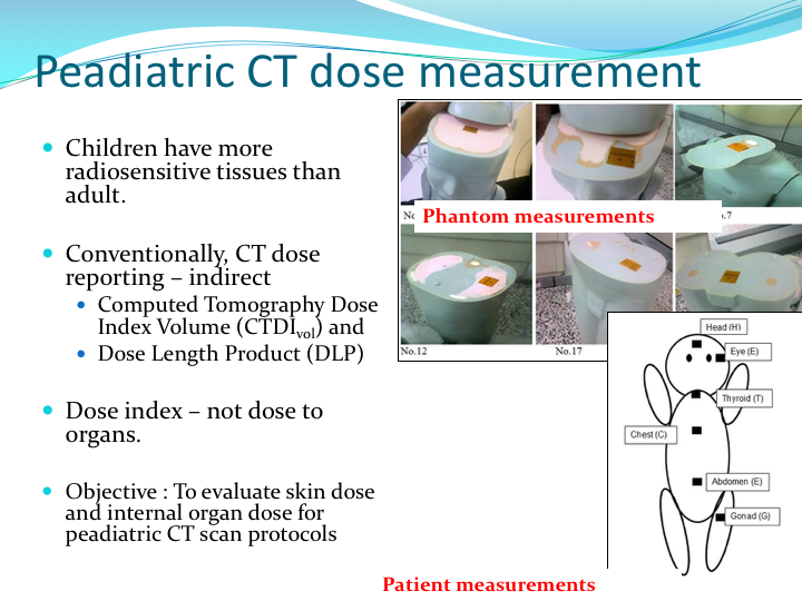
Peadiatric CT dose measurements
Children have more radiosensitive tissues than adult. Conventionally, CT dose reporting – indirect Computed Tomography Dose Index Volume (CTDIvol) and Dose Length Product (DLP) Dose index – not dose to organs. Objective : To evaluate skin dose and internal organ dose for peadiatric CT scan protocols
|
|
|
Validation of dosimetry system for high-dose rate (HDR) brachytherapy
To validate the HDR brachytherapy dosimetry system for in-vivo dose measurement. To measure rectal/bladder dose during brachytherapy and compare the results with TPS calculation.
|
|
|
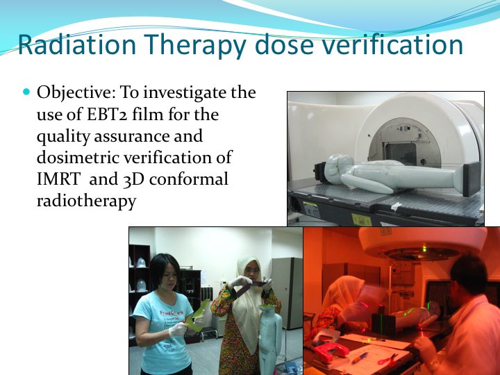
Radiochromic film dosimetry |
|
|
|
|
|
Research Team: Dr. Jeannie Wong Hsiu Ding Prof. Dr. Ng Kwan Hoong Dr. Ung Ngie Min Dr. Yeong Chai Hong Dr. Vincent Phua Chee Ee Mr. Shahrun Nizam Miss Khadijah Ramli Dr. Mohammad Amir Radhi bin Othman Prof. Dr. Anatoly Rosenfeld, Dr. Michael Lerch, Dr. Marco Petesseca Mohammad Javed Safari, Hanum Farsihah
|
|
|
Grant: UM.C/625/1/HIR/MOHE/MED38 RG507-13HTM
--- Updated: 25/9/2017 |
|